
Enzyme science · Deep dive
Catalase: structure, mechanism and industrial uses
Conversion of peroxide into water and oxygen allows residual peroxide removal after bleaching, cleaning and selected food-processing operations.
5 minute read
Recommended products →What is Catalase?
Catalase converts hydrogen peroxide into water and oxygen. Protects cells from damaging peroxide accumulation.
Catalase decomposes hydrogen peroxide into water and oxygen. It is central to oxidative-stress biology and useful where residual peroxide must be removed from a process.
Catalases protect cells by rapidly removing hydrogen peroxide generated during oxidative metabolism.
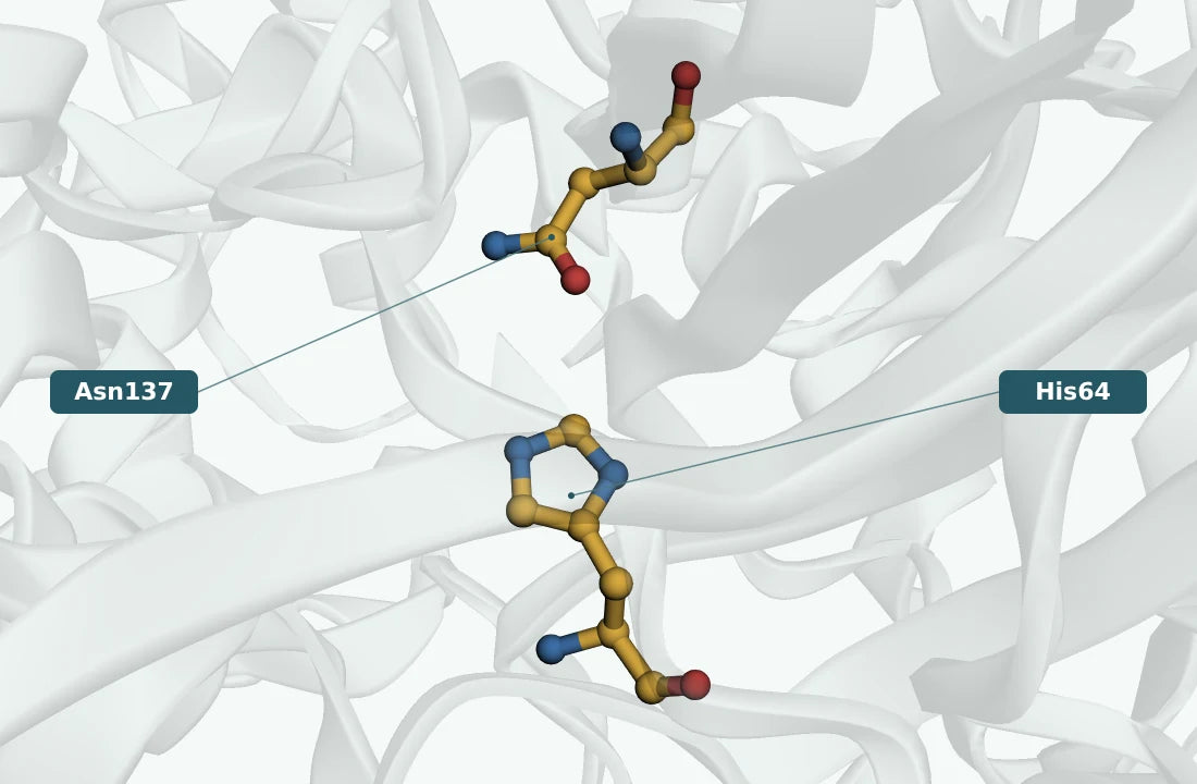
[3]The reaction scheme below connects starting materials and products; the active-site and energy diagrams explain the catalytic chemistry.

Essential properties and enzyme identity
| Property | Scientific detail |
|---|---|
| EC classification | EC 1.11.1.6 |
| Starting materials | Aqueous hydrogen peroxide remaining in a process stream or introduced into an analytical assay. |
| Products | 2 H₂O + O₂ |
| Representative molecular mass | About 75.6 kDa per deposited polypeptide for Catalase from Penicillium janthinellum (PDB 2IUF). |
| Subunit organisation | Homotetramer (4 subunits) for the named structural example. |
| Natural sources and production | Fungal and bacterial fermentation are established production routes; animal-derived enzymes also exist. |
| Key catalytic or process feature | Typical industrial examples are haem enzymes; subunit sizes and peroxide tolerance vary between catalases. |
Discovery and scientific milestones
Early biochemical evidence
Herbert and Pinsent reported crystalline bacterial and human erythrocyte catalases in 1948. Later structural work explained how haem intermediates support rapid peroxide disproportionation.
[3]Alfonso-Prieto and colleagues: molecular characterisation
The 2007 structural study resolved Catalase from Penicillium janthinellum, providing an experimental basis for examining its active site and substrate recognition.
[2]Published research milestone
Herbert, D. and Pinsent, J. Crystalline bacterial catalase. Biochem. J. 43 (1948) 193-202.
Original source ↗From natural sources to enzyme production
Catalases occur in animals, plants, fungi and bacteria. The structural example is a Penicillium enzyme historically studied as Penicillium vitale and recorded as Penicillium janthinellum.
Conventional production: Conventional microbial production uses controlled submerged fermentation in aerated vessels, with batch or fed-batch operation chosen for the producing strain. Secreted enzyme is recovered from clarified broth; intracellular production requires cell recovery and disruption. Concentration and formulation follow purification. Fungal solid-state cultivation is an alternative for suitable strains.
[1]
Reaction mechanism and active site
Typical haem catalases cycle through an oxidised intermediate called compound I. One peroxide molecule oxidises the haem system and another reduces it, releasing oxygen. Large-subunit fungal catalases and smaller catalases differ in architecture; non-haem manganese catalases also exist. The fungal model illustrates a tetrameric haem enzyme, not a universal form of every catalase.
- 1
One peroxide molecule oxidises the haem system
- 2
A second peroxide reduces the oxidised intermediate
- 3
Water and oxygen are released
Typical industrial examples are haem enzymes; subunit sizes and peroxide tolerance vary between catalases.
[2][3]Kinetics and catalytic performance
Hydrogen peroxide is both the reacting substrate and, at high exposure, a source of inactivation. Report the peroxide range and initial-rate window; a simple Michaelis–Menten fit may not describe the full range.
| Metric | Published range / example | Meaning and practical use |
|---|---|---|
| Kₘ | 0.125 mM | Substrate concentration at half Vmax for Michaelis–Menten kinetics; retain a polymer mass basis when used. |
| kcat | No matched value included. | Turnover per active catalytic centre at saturation; Vmax divided by active-site concentration. |
| kcat/Kₘ | No matched value included. | Low-substrate catalytic efficiency; compare the same substrate and conditions. |
| Vmax | 2,500 U/mL | Saturation rate for the stated enzyme loading; a protein-normalised value is identified by its units. |
| Specific activity | Assay- and loading-dependent; no intrinsic range. | Activity per mg protein under the specified assay; not necessarily a saturation rate. |
| v₀ | Assay- and loading-dependent; no intrinsic range. | Initial rate at the tested concentrations; changes with enzyme and substrate loading. |
Activity units: from measurement to useful conversion
| Measurement | Meaning |
|---|---|
| Typical activity definition | A common catalase unit represents decomposition of 1 μmol of hydrogen peroxide per minute. A Sigma assay example specifies pH 7.0, 25°C and 50 mM starting peroxide. |
| What the process measurement reveals | Residual peroxide is the process endpoint. Oxygen bubbles demonstrate reaction but do not quantify completion, and high peroxide exposure can affect enzyme survival. |
| U/g and U/mL | Activity per gram or millilitre of the supplied preparation. Specific activity in U/mg protein uses a different denominator. |
Industrial applications and research opportunities
Textile and other peroxide-based processes use catalase for residual peroxide removal. Food-processing and analytical systems may pair catalase with oxidases. Oxygen evolution can affect mixing and sampling, so disappearance of peroxide is the direct process endpoint.
 01
01Peroxide removal
Reduce residual peroxide after a process step.
Measure success: Measure peroxide directly to the required endpoint.
02Oxidase coupling
Manage generated peroxide.
Measure success: Account for oxygen returned by catalase.
03Enzyme research
Study rapid peroxide turnover and stability.
Measure success: Use a method with adequate time resolution.
Common questions
Does catalase require another organic electron donor?
In the typical catalatic reaction, peroxide serves both roles.
Do bubbles prove complete peroxide removal?
No. Gas evolution does not quantify the residual peroxide.
Are all catalases haem tetramers?
No. The displayed fungal enzyme is one defined example within broader catalase chemistry.
References and supporting evidence (7)
- RCSB PDB 2IUF: experimental coordinates, source and assembly
Protein-entity mass, coordinate model, experimental method and deposited biological assembly; checked 23 September 2026.
- Alfonso-Prieto, M.; Borovik, A.; Carpena, X.; Murshudov, G.; Melik-Adamyan, W.; Fita, I.; Rovira, C.; Loewen, P.C. (2007). The Structures and Electronic Configuration of Compound I Intermediates of Helicobacter Pylori and Penicillium Vitale Catalases Determined by X-Ray Crystallography and Qm/Mm Density Functional Theory Calculations.
J.Am.Chem.Soc. 129:4193–. Primary experimental structural publication.
- IUBMB enzyme nomenclature: EC 1.11.1.6
Accepted reaction, classification and historical bibliography.
- UniProt D9N167: protein annotation
Curated protein identity and available subunit annotation;
- Herbert, D. and Pinsent, J. Crystalline bacterial catalase. Biochem. J. 43 (1948) 193-202.
Primary study listed in the IUBMB nomenclature bibliography.
- Herbert, D. and Pinsent, J. Crystalline human erythrocyte catalase. Biochem. J. 43 (1948) 203-205.
Primary study listed in the IUBMB nomenclature bibliography.
- Sigma-Aldrich: catalase activity definition
Assay substrate, reporting convention and reference conditions.
Recommended products
Choose the products that match your process. Each card explains its role in this application; you do not need every enzyme in one recipe.

Catalase
From £78.99
View sizes & buy
Catalase (powder)
From £178.99
View sizes & buyBenefits are application targets; confirm dosage and performance in your finished formulation.