
Enzyme science · Deep dive
Keratinase: structure, mechanism and industrial uses
Keratin breakdown can support feather-waste valorisation, peptide recovery and development of enzymatic dehairing processes.
5 minute read
Recommended products →What is Keratinase?
Keratinase degrades accessible keratin proteins. Recycles nitrogen from feathers, hair and related materials.
Keratinase denotes proteolytic activity against resistant keratin-rich materials. It is a functional category encompassing different enzymes, rather than one protein with a universal structure.
Keratinolytic microorganisms recycle recalcitrant proteins in feathers, hair and other keratin-rich biological materials.
[3]The reaction scheme below connects starting materials and products; the active-site and energy diagrams explain the catalytic chemistry.

Essential properties and enzyme identity
| Property | Scientific detail |
|---|---|
| EC classification | No single EC number covers every keratinolytic enzyme; EC 3.4.21.62 describes subtilisin-type serine proteases, including the representative family discussed here. |
| Starting materials | Feathers, hair, wool and prepared keratin substrates with different degrees of physical accessibility. |
| Products | Keratin-derived peptides |
| Representative molecular mass | About 72.0 kDa for the deposited fervidolysin precursor construct (PDB 1R6V); processing produces the mature enzyme. Keratinase preparations span multiple protease families. |
| Subunit organisation | Monomer for the named structural example. |
| Natural sources and production | Keratinolytic Bacillus strains and other bacteria or fungi; Fervidobacterium pennivorans supplies the thermophilic structural example. |
| Key catalytic or process feature | Disulfide crosslinks and compact fibre architecture restrict access; peptide cleavage and structural opening are complementary processes. |
Discovery and scientific milestones
Early biochemical evidence
Keratin-degrading activity has been investigated in different proteases and organisms. The structural study below characterises fervidolysin and its precursor-related architecture. It is evidence about one named enzyme, not a claim that all keratinases were discovered in that year or share its molecular mass.
[3]Kim and colleagues: molecular characterisation
The 2004 structural study resolved Subtilisin-like serine protease from Fervidobacterium pennivorans, providing an experimental basis for examining its active site and substrate recognition.
[2]Feather-waste valorisation
Bacillus pacificus RSA27 keratinase was investigated for converting feather waste into protein hydrolysate.
Published source ↗From natural sources to enzyme production
Keratin-degrading Bacillus strains and fungi are widely studied. Fervidobacterium pennivorans provides a thermophilic example with a different ecology and architecture from many mesophilic enzymes.
Conventional production: Conventional microbial production uses controlled submerged fermentation in aerated vessels, with batch or fed-batch operation chosen for the producing strain. Secreted enzyme is recovered from clarified broth; intracellular production requires cell recovery and disruption. Concentration and formulation follow purification. Fungal solid-state cultivation is an alternative for suitable strains.
[1]
Reaction mechanism and active site
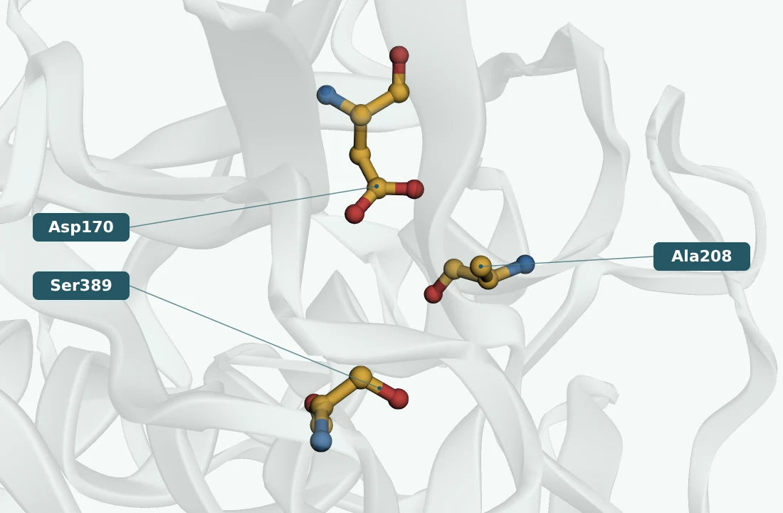
Keratin packing and disulfide crosslinks limit access. Proteases cleave peptide bonds; disruption of disulfide-stabilised structure can be a separate enabling step. Fervidolysin, the illustrated example, is a subtilisin-related serine protease. Maturation removes precursor regions, changing the mass of the active protease.
The native subtilisin-like catalytic system uses Asp170, His208 and Ser389 in the author numbering of this structure. The displayed 1R6V model is an H208A precursor mutant: Ala208 marks the substituted histidine position, rather than an intact catalytic triad.
- 1
Expose susceptible regions in the keratin material
- 2
Hydrolyse accessible peptide bonds
- 3
Recover and characterise soluble peptides and remaining solids
Disulfide crosslinks and compact fibre architecture restrict access; peptide cleavage and structural opening are complementary processes.
[2][3]Kinetics and catalytic performance
Keratin is insoluble and crosslinked. Particle size, disulfide reduction and pretreatment can dominate hydrolysis, so keratin mass loss and soluble peptide release complement activity on soluble proteins.
| Metric | Published range / example | Meaning and practical use |
|---|---|---|
| Kₘ | 9.8 mg/mL | Substrate concentration at half Vmax for Michaelis–Menten kinetics; retain a polymer mass basis when used. |
| kcat | No matched value included. | Turnover per active catalytic centre at saturation; Vmax divided by active-site concentration. |
| kcat/Kₘ | No matched value included. | Low-substrate catalytic efficiency; compare the same substrate and conditions. |
| Vmax | 307.7 µmol/min | Saturation rate for the stated enzyme loading; a protein-normalised value is identified by its units. |
| Specific activity | Assay- and loading-dependent; no intrinsic range. | Activity per mg protein under the specified assay; not necessarily a saturation rate. |
| v₀ | Assay- and loading-dependent; no intrinsic range. | Initial rate at the tested concentrations; changes with enzyme and substrate loading. |
Activity units: from measurement to useful conversion
| Measurement | Meaning |
|---|---|
| Typical activity definition | Keratinase units are strongly method-dependent. Keratin azure and azokeratin methods often define activity through dye release or absorbance change per minute; soluble-protein protease units are not equivalent. |
| What the process measurement reveals | Loss of insoluble keratin, soluble peptide formation and peptide size are useful conversion measures. Activity on casein does not establish degradation of intact feathers. |
| U/g and U/mL | Activity per gram or millilitre of the supplied preparation. Specific activity in U/mg protein uses a different denominator. |
Industrial applications and research opportunities
Applications under development include feather-waste valorisation, keratin peptide production and selected leather processes. Product quality, digestibility and mass balance matter alongside disappearance of starting material.
 01
01Feather valorisation
Generate useful protein-derived fractions.
Measure success: Report recovered peptides and residual mass.
02Keratin materials
Investigate controlled modification.
Measure success: Preserve the required material properties.
 03
03Leather research
Explore selective treatment of protein structures.
Measure success: Measure effects on both target and retained components.
Common questions
Is keratinase a single catalytic family?
No. The term describes activity on keratin and includes different proteases.
Does a casein assay predict feather digestion?
Not reliably; keratin presents additional accessibility barriers.
Why is the displayed mass not a universal keratinase mass?
The research construct and maturation state are specific to that protein.
References and supporting evidence (5)
- RCSB PDB 1R6V: experimental coordinates, source and assembly
Protein-entity mass, coordinate model, experimental method and deposited biological assembly; checked 23 September 2026.
- Kim, J.S.; Kluskens, L.D.; de Vos, W.M.; Huber, R.; van der Oost, J. (2004). Crystal structure of fervidolysin from Fervidobacterium pennivorans, a keratinolytic enzyme related to subtilisin.
J.Mol.Biol. 335:787–797. Primary experimental structural publication.
- IUBMB enzyme nomenclature: EC 3.4.21.62
Accepted reaction, classification and historical bibliography.
- UniProt Q93LQ6: protein annotation
Curated protein identity and available subunit annotation;
- Sigma-Aldrich: keratinase activity assay
Assay substrate, reporting convention and reference conditions.
Recommended products
Choose the products that match your process. Each card explains its role in this application; you do not need every enzyme in one recipe.

Keratinase
From £78.99
View sizes & buyBenefits are application targets; confirm dosage and performance in your finished formulation.